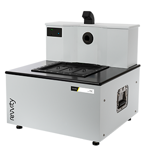
Vegaは、Revvity社が誇る最先端の前臨床生体イメージング技術を搭載した最新システムです。
革新的な設計のVegaは、わずか数分で高解像度の2Dおよび3D画像を取得可能なハンズフリーの自動超音波プラットフォームです。
ハンズフリー、自動化、高速・広視野
Vegaは、基礎研究において超⾳波を簡単に利⽤できるように設計された新しいコンセプトに基づくシステムで、従来の⼿持ち型超⾳波プローブ特有の様々な課題を克服します。
イメージングステージの下部に設置された⾃動トランスデュー サにより、ハンズフリーによる超⾳波イメージングはわずか数分で完了します。専任のテクニシャンは不要で、観察したい部位を正確に⻑期間観察することが可能です。
Vega Ultrasound Imaging System from Revvity
特長
- ハンズフリー -トランスデューサーの自動配置と移動
- 最小限のトレーニングで簡単に使用可能
- わずか数分で3匹のマウスをスキャンできる高速、高スループットのパフォーマンス
- 3D広視野取得により、対象全体のイメージングが可能
- 標準BモードおよびMモード機能
- 組織の硬さを定量化するせん断波エラストグラフィー(SWE)モード
- 微小血管を視覚化する音響血管造影(AA)モード
- 柔軟な視覚化および分析ソフトウェア
- ベンチトップに収まる
Vega Innovator Contest
機能

ハンズフリー
従来の手持ち型プローブの限界を克服するハンズフリー超音波イメージングにより、一貫した結果を得ることが可能

自動化
測定ステージ下に配置された自動トランスデューサによる下方からのイメージングは、簡単に使用できるため専任の超音波検査技師を必要としません

ハイスループット
測定はわずか1分です。3匹のマウスの連続測定と効率的なワークフローにより、超音波イメージンのスループットを向上します。

広視野超音波
高速3D全身測定で、疾患や治療効果を広い解剖学的視点で可視化します。

多様な用途に対応する
多機能モード
2種類の統合型トランスデューサにより、高解像度・血管イメージングから深部組織、エラストグラフィー、心臓イメージングまで幅広い用途に対応

エラストグラフィー
シアウェーブエラストグラフィー(SWE)で数秒で組織の硬さを評価・測定し、疾患の進行を確認できます。

造影超音波(CEUS)
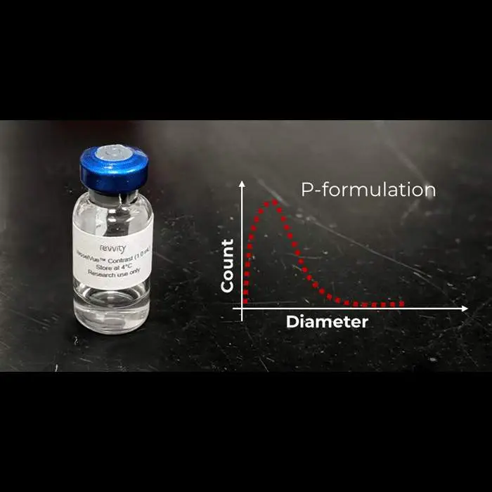
Acoustic AngiographyモードとVesselVueマイクロバブル造影剤で微小血管を可視化し、腫瘍血管網や密度の定量化、治療効果や組織損傷の経時的変化を確認します。

柔軟性
Vegaはイメージングステージ下に2つのトランスデューサを内蔵し、手動での交換が不要で、モード切り替えが簡単で柔軟に行えます。
資料
資料のダウンロードには、ユーザー登録が必要です。
登録がお済みでないお客様は、こちらからユーザー登録をお願いいたします。
Recent advances in noninvasive imaging technologies have introduced innovative approaches to longitudinally track changes in...
Researchers at the University of North Carolina use automated, high throughput ultrasound and acoustic angiography as an...
Being able to characterize and monitor the extent of fibrosis, steatosis, and inflammation in the liver non-invasively is valuable...
Researchers trust our in vivo imaging solutions to give them reliable, calibrated data that reveals pathway characterization and...
The need for better profiling is clear, and technological advances are making it possible to optimize clinical trials and...
Given the prevalence of fibrosis in chronic liver diseases, numerous therapeutic approaches for managing liver disease prioritize...
The primary goal of preclinical imaging is to improve the odds of clinical success and reduce drug discovery and development time...
Monitoring tumor response to therapy typically relies on changes in tumor volume. However, various functional and molecular...
動画
関連製品
VesselVueマイクロバブル造影剤で血管を可視化