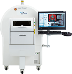
CosmoScanは、「高速撮影」「高解像度」「広視野」「低被曝」を実現した、生体X線イメージングに特化した実験動物用3DマイクロX線装置です。
5ミクロンの高解像度と、8~86mmの広視野を実現し、独自のアクティブリング除去や高度な呼吸ゲーティングにより、連続・ステップ走査にも対応します。更に光イメージング装置であるIVIS Imaging Systemとのシームレスな画像融合も可能です。
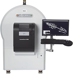
CosmoScan GX IIIは、マウス・ラットから小型のウサギまで、高速・高分解・広視野で撮影可能な実験動物用3DマイクロX線CTの最新・最上位機種です。 最速3.9秒の超高速撮影、最高5μmの超高分解能撮影、最大FOV86×240mmの超広視野撮影が可能です。
CosmoScan GX IIIは、実験動物用CTに求められる三大要素(高速・高分解能・広視野)を業界最高レベルで実現したプレミアムCTです。GX IIIを用いることで、In vivoからex vivoまで1台で多様なアプリケーションにご利用頂くことが可能です。
2006年から当社主催の「In vivo imaging form」を毎年開催しており、2025年10月には第18回目*1を迎えました。本ユーザー会には、光イメージング装置IVISユーザー様の他、CosmoScan CTをご利用頂いている方にも多数ご参加頂き、貴重な情報交換の場としてご活用いただいております。
ユーザー会では、ユーザー様による貴重な講演に加え、学生を中心としたポスター発表などの企画も実施。毎年、多くのリピーターの皆様にもご参加いただいております。アットホームでフレンドリーな雰囲気の会ですので、ぜひお気軽にご参加ください。
*1 2020年、21年はコロナ禍により未開催。
Resolution that’s remarkable! The Quantum*2 GX3 microCT preclinical imaging system
*2 リガク社CosmoScan GX IIIの海外版です。
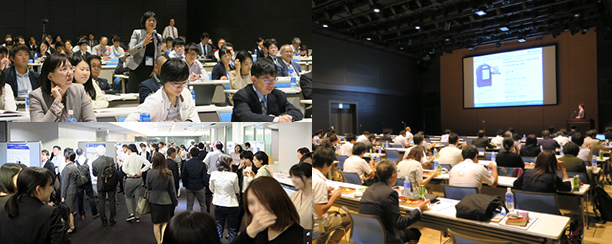
弊社主催 In vivo imaging forumの様子
特長 - CosmoScan GX III -
- 最高空間分解能(5µm)
- 最高速スキャン(3.9秒)
- 高出力X線(20W)
- アクティブリングリダクションによるアーチファクトの自動除去と画質向上
- 5種類の視野範囲(8mmから86mm)
- ex vivoとin vivoのサンプル解析が可能
(マウスから小さなウサギまで) - 強化された画像ベースの2相心拍・呼吸同期
- 低線量撮影により長期的なモニタリングが可能
- ex vivoサンプルに最適なStep Scanモード
- IVISによる光イメージングとの簡単な画像融合
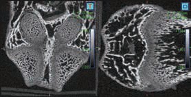
より詳細に解剖学的構造を視覚化
優れた空間分解能
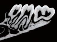
CosmoScan GX IIIは特に高い空間分解能を持ち、5µmまでの解像度と2.9µmのピクセルサイズ(FOV:8mm)を誇ります。これにより小さい骨の細かな解剖学的構造を含むex vivoサンプルの高品質な画像が得られます。
- 空間分解能 = 5µm 最小ピクセルサイズ = 2.9µm
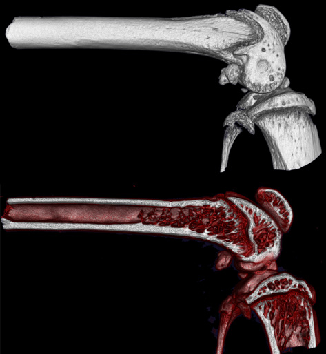
図1. マウスの膝の皮質(上)と梁状(下)の画像
強化されたX線
改良されたX線源(20〜100kV、20W)を搭載したGX IIIは、軟組織のような低密度のサンプルから骨のような高密度の物体まで、幅広いサンプルに対して高いX線透過性を実現しています。その結果、シグナル対ノイズ比が向上し、より優れた画像品質が得られます。
画像品質が向上したその他の方法
標準的なビームハードニング補正に加えて、独自の統合型Active Ring Reduction(ARR)ハードウェアを新しく搭載し、リング状のアーチファクトを自動的に取り除き、画像品質を向上させています。
このシステムはStep Scanモードを使用して動きによるアーチファクトを低減するとともに、画像ベースのゲーティング技術と組み合わせることで、業界最高レベルの画像品質と解像度を実現しています。
- 画像ベースの心拍・呼吸同期 - 肺や心臓の動きに起因するアーチファクトを低減
- Active Ring Reduction - リング状のアーチファクトを自動で除去
- Step Scanモード - 動きによるアーチファクト削減のための「ステップアンドシュート」
- 改善された透視機能 - 実時間での解剖学的構造や血管のより良い可視化
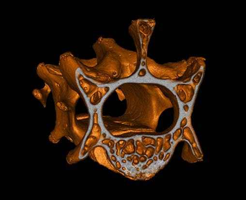
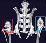
図2. マウスの椎骨の断面図
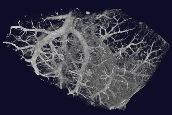
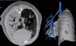
図3. マウスの肺のex vivoイメージング
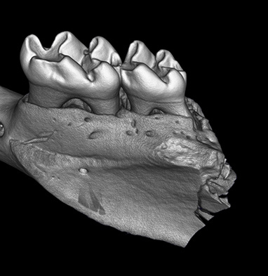
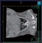
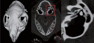
図4. マウスの顎
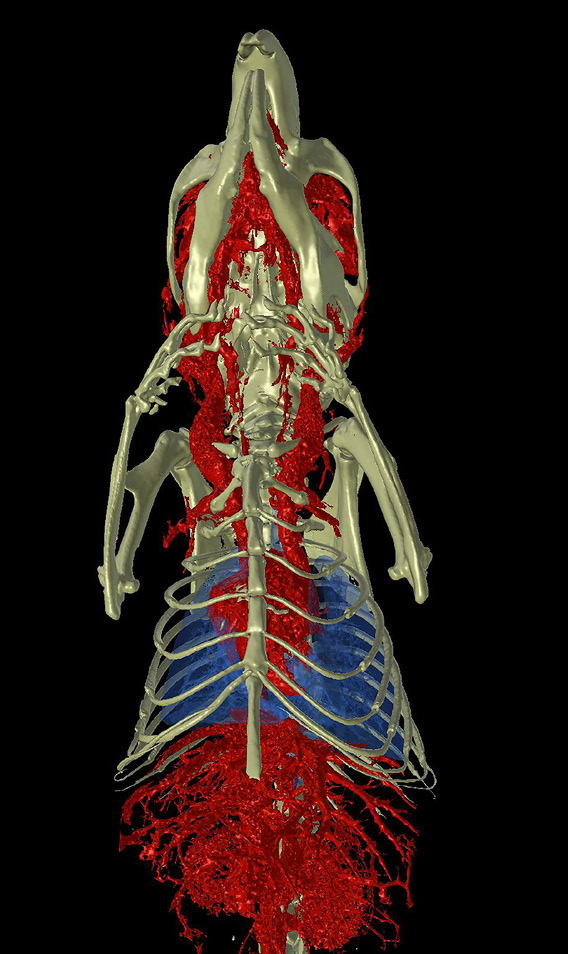
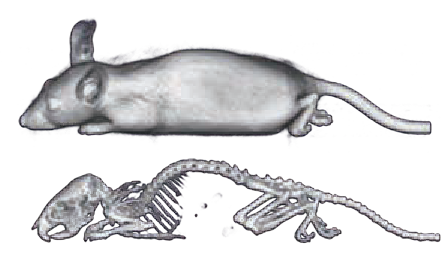
図5. 骨格、肺、および血管を示すマウス上半身画像
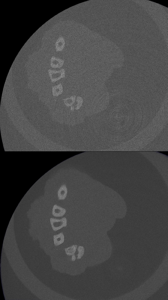
図6. Active Ring Reduction(ARR)の比較(上)ARR実装前(下)ARR実装後
優れた視野範囲(FOV)
GX IIIは、マウスから小型ウサギまでのさまざまな種のin vivoイメージングだけでなく、より高解像度なex vivo イメージングも可能となり、さまざまなサンプルをより最適な測定方法で撮影できる幅広い柔軟性を提供します。
- 5種のFOV:8mm, 18mm, 36mm, 72mm, 86mm
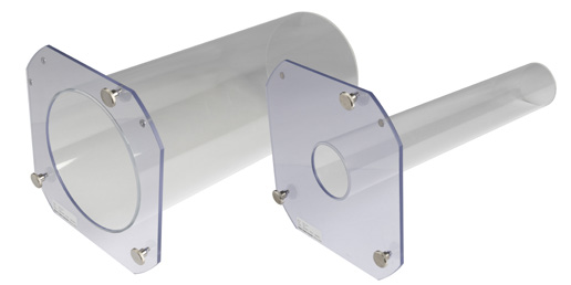
- ボアサイズ:163mm
- スキャン可能範囲は240mm
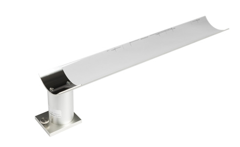
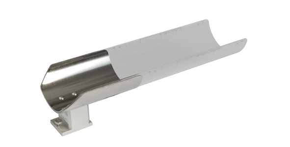
- Ex vivoサンプル用ホルダーは8mm FOVで使用可能
- 複数の種を収容するための動物用ベッド
FOV(視野)とサンプル種の例
| FOV | サンプルや対象物の例 |
|---|---|
| 8mm | Ex vivoサンプル |
| 18mm | ゼブラフィッシュやその他の小さなサンプル |
| 36mm | 標準的なマウスイメージング |
| 72mm | ラットやウサギ |
| 86mm | 大型動物の肺を1回のスキャンで撮影 |
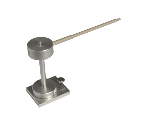
図7. 8mm視野範囲のサンプルホルダー

図8. マウス椎骨のex vivo画像
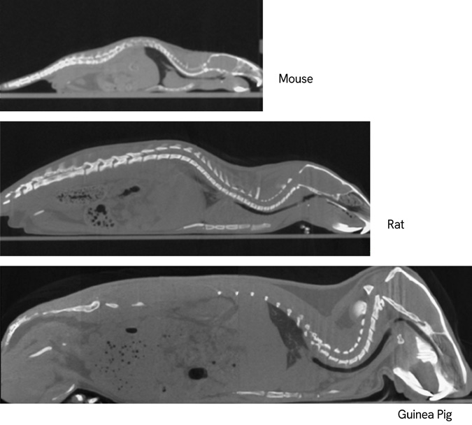
図9. 幅広い生物サンプルや動物種に対応するための、調節可能な視野範囲、大きなボアサイズ、広いスキャン範囲
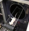
マウス・ラット・フェレット対応
2相心拍・呼吸同期CT
心拍や呼吸による動きは、CT画像のアーティファクトの原因となります。GX IIIは画像ベースの同期技術により、マウス、ラット、フェレットの心臓・肺イメージングを高精度に実現します。
撮影後、心臓や横隔膜に設定したROIをもとに独自アルゴリズムで同期処理を行い、アーティファクトを低減。心拍・呼吸周期の特定フェーズのデータを再構成することで、画質と定量精度を向上させます。
心臓・肺のin vivoイメージングなど、動きの影響を受けやすい研究に最適です。
GX IIIは高速モードにより、スキャン時間わずか3.9秒を実現します。再構成も約6秒で完了し、最短約10秒以内に3D CT画像の取得から再構成までを行うことが可能です。
この高速性能により、長期研究における疾患の進行を効率的かつ容易に追跡・解析できます。
3.9秒の高速スキャンにより、良好な画質と低X線量を両立したCT撮影が可能です。これにより、疾患モデルの長期的な評価を無理なく実施できます。
また、高速撮影とスムーズなワークフローにより、多数のサンプルを効率的に撮影し、実験データから正確な結論を迅速に導出できます。
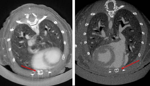
図10. マウス肺の画像ベースゲーティング:(左)ゲーティング前、(右)ゲーティング後。赤い矢印は、胸骨におけるモーションアーチファクトの除去によるゲーティング後の画像品質の向上を示す。
連続スキャン/ステップスキャンモード
高速・高スループットな連続スキャンモードと、高解像度撮影に適したステップスキャンモードのいずれかを用途に応じて選択できます。
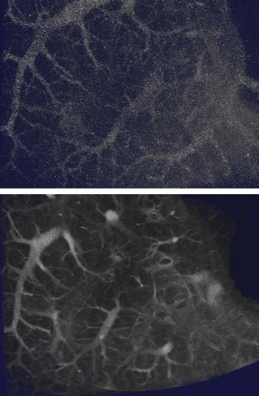
図11. 8mm FOVを用いた肺標本のEx vivo撮影(上)連続スキャン、(下)Step Scanモード
自動フィルター検知機能
CosmoScan GX IIIは、低エネルギーX線を除去してX線品質を向上させる6種類のフィルターを搭載しています。
スキャン開始前に、適切なフィルターが選択されているかを自動で検知・確認し、安定した撮影をサポートします。
| フィルター (材質の厚み) | アプリケーション例 |
|---|---|
| Al 0.5mm | 低コントラストサンプル (in vitroの脳など) |
| Al 1.0mm | 軟組織のスキャン (脂肪の分析など) |
| Al 0.5mm + Cu 0.06mm | 標準的なCTスキャン |
| Cu 0.1mm | 高電圧時の高密度サンプル |
| Cu 0.2mm | 金属を含むサンプル (歯科インプラントなど) |
| Cu 1.0mm | X線発生器のウォームアップ時の フラットパネル検出器の保護 |
GX IIIは、イメージングの最適化のために6つの交換フィルターと、フィルター自動感知機能を搭載しています。
光イメージングとの融合
マイクロCTを他のモダリティと組み合わせることで、生体の健康状態や疾患の構造的・機能的理解が深まります。
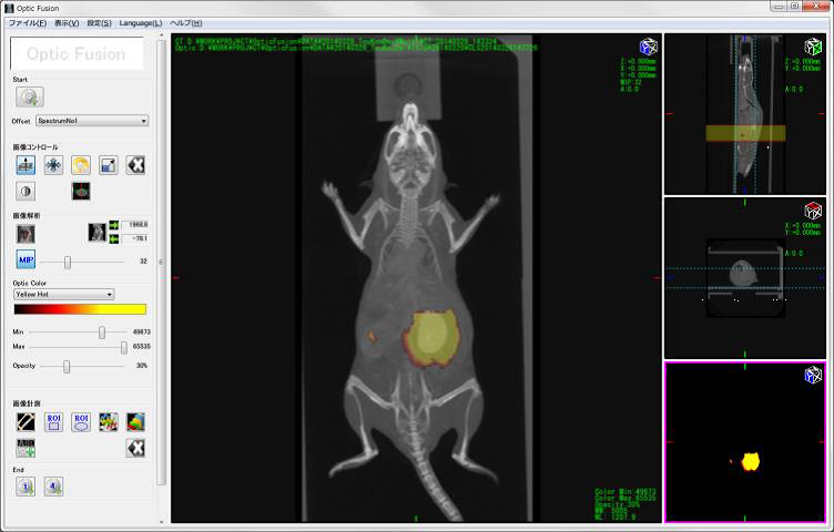
GX IIIは、専用のマウスイメージングシャトル(MIS)を用いて、マイクロCTデータとIVIS Imaging Systemのデータをシームレスに統合可能です。シャトルは両プラットフォームに容易に装着でき、撮影中の位置再現性を維持します。
基準マーカー機能により、数クリックで自動データ融合を実現。取得データはDICOM形式でエクスポートでき、各種解析ソフトウェアで利用できます。
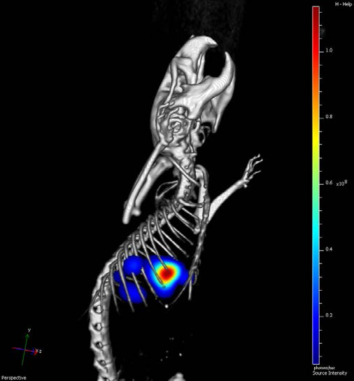
図12. マウスの肺に移植されたIVISense腫瘍細胞株のIVIS Spectrumによる光イメージングとGX IIIによるCT画像の融合
ソフトウェアの主な特長
- 迅速でシームレスな解析 -
CosmoScan GX IIIは、直感的で使いやすい操作性により、データ取得から解析までスムーズに行えます。
スキャン直後に画像再構成が自動で開始され、基本ツールを用いて組織密度の可視化や数値測定、詳細解析用データの出力を簡単に実施できます。
- マイクロCTデータの取得・閲覧を直感的に行える操作性
- 距離測定やROI面積測定に対応した解析ツール
- 閾値設定による鮮明な3D表示
- スタディスキャンを階層的に管理できるイメージデータベース
- ジョブスキャン機能による自動連続撮影やパノラマ画像作成
- サブボリューム再構築による高解像度イメージング
- IVISの3D光イメージング画像との容易な融合
- サブボリューム再構築による高解像度イメージング
- DICOM形式でのデータエクスポートに対応し、他ソフトでの追加解析が可能
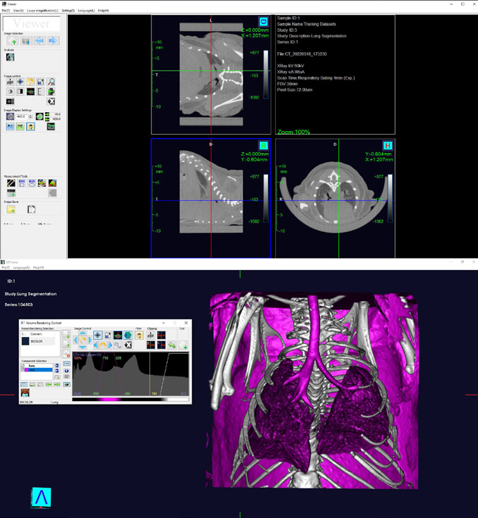
図13. 視覚化と解析のための使いやすいソフトウェア。Viewerソフトウェアによる終呼吸時のマウス肺の2D断面図(上)と3Dレンダリング(下)。
Analyze 14.0 ソフトウェア(オプション)
Analyze 14.0ソフトウェアを用いることで、骨、体組成、脂肪量などのより高度な解析が可能になります。
- 解剖領域を高精度かつ迅速にセグメンテーション
- 長さ・体積・強度測定による詳細な統計解析
- フィルタリングによる最適な画像可視化
- マイクロCTデータと他モダリティ画像の重ね合わせ解析
- 高解像度の静止画および3D動画の表示・保存
- 空間変換、輝度変換、数学的処理、データ再配置に対応
- 骨微細構造解析(BMA)機能(オプション)
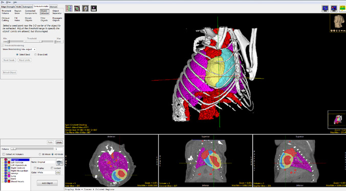
図14. Analyze 14のセグメントモジュールに含まれる、組織や領域の迅速かつ正確な定義のための高度なインタラクティブツール
CosmoScan GX II
ソフトウェア最新バージョン(3.3.0)主な追加機能
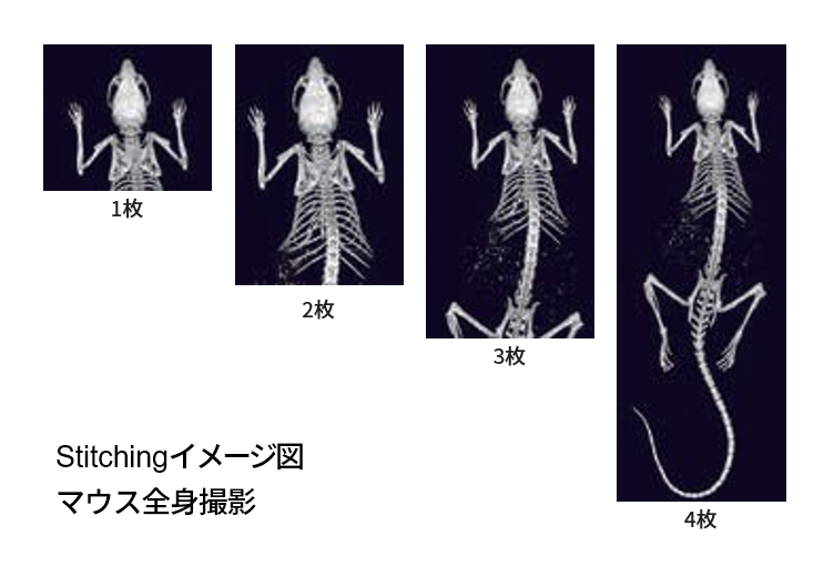
高解像度画像 Stitching 機能
- 全視野での高解像度画像の結合が可能
- 画像表示領域を大幅に拡大*3
*3 従来のWhole Body Scan Z軸方向最大
120mm → 240mm
Job Scan 機能
- 複数のサンプルを連続で撮影
- サンプルごとの撮影条件設定が可能
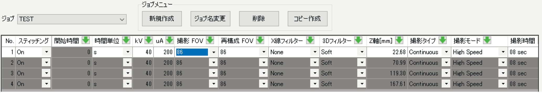
Job Scan 設定画面
CT Image Resolution 選択機能
- 1K-1K再構成画像表示・出力機能を搭載
- 目的に応じた画像出力サイズを選択可能
対象機種
- CosmoScan GX II*4
- CosmoScan FX, GX*5
*4 GPU交換が必要な場合があります。別途ご相談ください。
*5 HDDの増設やPC、GPU交換が必要な場合があります。
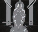
マウス頭部撮影 1K×1K再構成画像
その他の機能改善
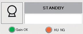
- 各種校正ステータス表示
校正ステータスをソフトウェア上で容易に確認 - 画像回転表示時の画像高解像度化
Viewerで回転表示した画像の高解像度化(再々構成)が可能
GainおよびHU校正のステータス
画像回転時の再々構成範囲指定
画像回転前
画像回転後
高速撮影モード
- 最新の超高速検出器搭載により最速3.9秒スキャン実現
- 高速画像再構成約10秒を実現
- サンプルの被ばくを最小限に低減
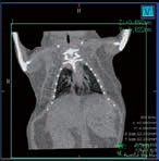
呼吸リズムが不規則な個体の肺野撮影
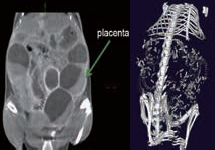
妊娠マウス
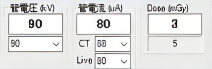
被ばく線量表示機能搭載
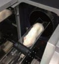
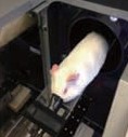
広視野撮影モード
- 5kgウサギの撮影にも対応。FOV86を実現
- 肥満モデル動物の心拍呼吸同期にも対応
Nude mouse(25g)
Rat(220g)
Guinea pig(650g)
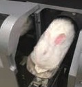
Rabbit(5kg)
ラット心拍呼吸同期撮影(ライブモード)
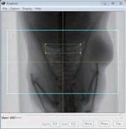
Super High Resolution モード
- 超接写イメージングが可能
- 最小解像度2.3μmを実現
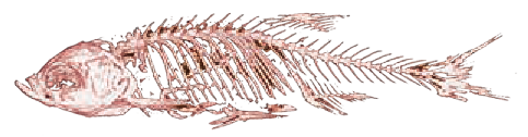
ゼブラフィッシュ骨格
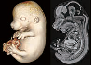
マウス胎児
マウス大腿骨
マウス顎骨
CosmoScan GX
- 最速8秒撮影、最速10秒画像再構成
- 最大FOV72mm撮影モード搭載
- 最高解像度4.5μm*6
*6 FOV36、画像再々構成機能使用時の最高解像度。
画像再々構成機能はGX/GX IIのみ搭載可能
FOV72mm High resolution scan Fine structure in the inner ear

4.5µm
FOV45mm

4.5µm
CosmoScan FX
- 最速18秒撮影、高速画像再構成最速10秒
- 最大FOV72mm撮影モード搭載
- 最高解像度10μm
- 生体監視用ライブモード
- マウス・ラット用の心拍呼吸同期撮影機能
- 奥行最大120mm ホールボディスキャンモード
- 脂肪解析ソフトウェア
ホールボディスキャン
オプション
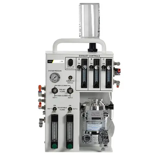
RAS-4 Rodent Anesthesia System
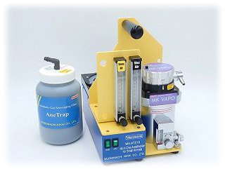
オールインワン小動物用麻酔器
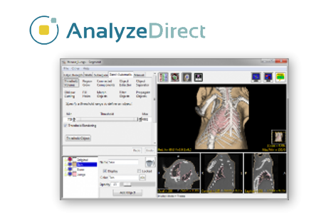
Analyze 14.0 ソフトウェア

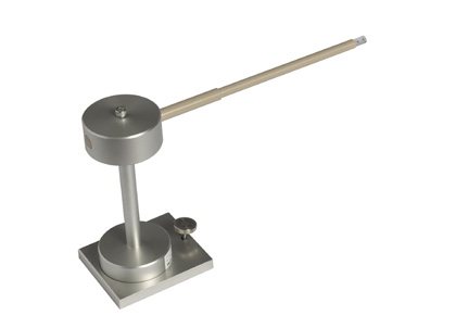
マウス頭部固定具
MIS Mouse Imaging Shuttle KitIVIS Spectrumシリーズとの3D画像融合や、Luminaシリーズの2D画像融合に使用
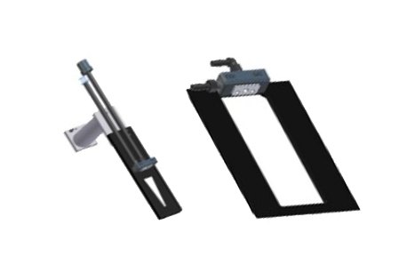
CT Arm AdaptorMISキットをCTで使用するためのキット
CT-Optic Fusion KitIVIS Luminaシリーズとの2D画像融合が可能になるキット
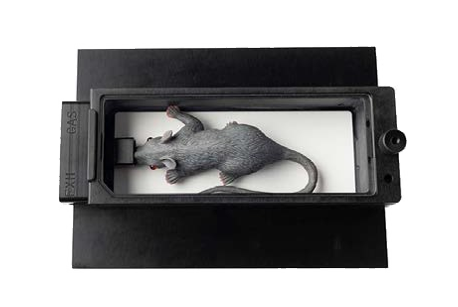
8mm FOV試料ホルダー
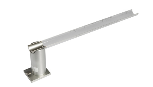
小型げっ歯類用のベッド
大型げっ歯類用のベッド
ウサギ用のベッド
ボアカバー: 70Φ·170Φ
機種別比較表
| スペック比較 |  |
|
 |
|||
|---|---|---|---|---|---|---|
| 機種名 | GX III | GX II | GX | FX | AX | |
| 主な用途 | Ex vivoから 小型実験動物まで |
小型実験動物から ex vivoまで |
小型実験動物向け | 中型動物向け 小型のウサギから 15kg程度まで |
||
| 主な性能 | 最高空間分解能 | 5µm | 15µm | 20µm | 60µm | |
| 最大管電圧 | 100kV | 90kV | 80kV | |||
| X線管球 | 最大管電流 | 200µA | 500µA | |||
| 最大出力 | 20W | 8W | 40W | |||
| 検出器 | タイプ | フラットパネルディテクター | ||||
| Frame Rate | 119fps | 117fps | 60fps | 30fps | ||
| CTガントリ | Bore Size | 163mmφ | 322mmφ | |||
| Scannable Range | 240mm | 440mm | ||||
| CT画像 | 撮影視野(FOV) | 86mm | 72mm | 220mm | ||
| 最長撮影視野 (体軸方向) |
240mm | 120mm (スティッチ機能搭載で240mm) |
120mm | 130mm | ||
| ピクセルサイズ | 2.9µm(min.) | 2.3µm(min.) | 4.5µm(min.) | 10µm(min.) | 60µm(min.) | |
| 画像数 | 512 × 512 × 400~ 2864 × 2864 × 2272 |
512 × 512 × 400~ 8000 × 8000 × 4300 |
512 × 512 × 512 | |||
| 撮影時間 | 連続スキャン | 3.9秒~70分 | 3.9秒~57分 | 8秒~57分 | 18秒~4分 | 18秒~2.5分 |
| Step Scan (長時間撮影) |
有 | 無 | ||||
| ソフトウェア | リングアーチファクト 低減 |
アクティブ補正 ソフトウェア補正 |
ソフトウェア補正 | |||
| その他 | 本体の大きさ (H×W×D) |
1536 × 960 × 963mm | 1536 × 960 × 1295mm | |||
| 本体重量 | 530kg | 450kg | 580kg | |||