
![]()

MediLumineAngiofil
Vascupaint Silicone Rubber Injection Compounds
|
||||||||||||||||||||||||||||||||||||||||||||||||||||||||||||||||||||
| 詳細 | |
|---|---|
| 製品説明 | Vascupaintは微小血管の構造の観察を目的とした血管評価用試薬です。本製品はYellow(マイクロCT用)に標識されており、血管内に充填・組織を透明化処理することで、明視野顕微鏡、マイクロCTによる目視検査や3Dイメージングを可能にします。 |
| 特長 | ・Ex vivo用造影剤 ・従来製品である鉛ベースの化合物よりも粘度が低く、非生体や臓器への灌流が容易 ・血管系の3Dイメージングに最適 |
| 参考文献 | |


左画像:Vascupaint Yellow灌流後にSkyScan 1276イメージング・システム, 解像度:6ミクロンで撮影。Dr. Sean Marrelli (UT Health) and Bruker Preclinical Imaging (Billerica, MA)提供
右画像:Vascupaint Yellow灌流後のマウス脳血管の明視野顕微鏡画像。Dr. Sean Marelli, Department of Neurology, McGovern Medical School at UT Health 提供
| 製品名 | 容量 | 製品コード | 価格(税抜) |
|---|---|---|---|
| Vascupaint Yellow Kit | シリコンボトル:200mL 希釈剤:200mL 触媒:20mL |
MDL-121 | 96,000 |
Fenestra LC
| 詳細 | |
|---|---|
| 製品説明 | Fenestra LCは小動物の肝臓、脾臓観察用の造影剤です。肝細胞を選択的に標的とし、機能的、解剖学的イメージングや脂肪肝のステージ分類・モニタリング、また肝腫瘍の体積測定などにご使用いただけます。 |
| 特長 | Fenestra LCは最終的に代謝され肝胆道系から排出されるため、複数回投与でき継続的なモニタリングが可能 |
| 参考文献 | |
| アプリケーション ノート |
|
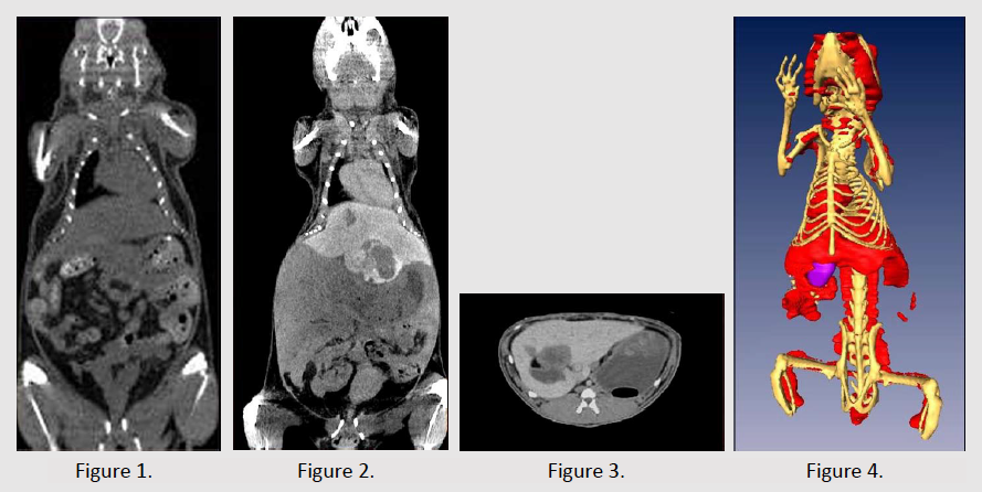
Fig1: 造影剤を使用せず撮影(マウス, オス, 腹腔側)。胸腔と腹腔で軟部組織のコントラストが悪い。
Fig2-4: Fenestra LC, iv投与2時間後のCT-26担癌マウス像(MicroCAT II撮影)
腹腔側: 冠状面(Fig2)、横断面(Fig3)、3D再構成(Fig4)
肝ドーム近くのX線吸収率の低い胆のうは腹腔側の画像(Fig2)で容易に確認できる。CT-26腫瘍は肝臓の下縁に観察され、腫瘍の下部にenhanced liverがあるのが確認された。CT-26腫瘍を3D再構築した画像がFig4であり紫色で表示した。
(詳細:![]() Imaging Hepatic CT 26 Tumors in BALB/c Mice as a Model of Metastatic Liver Cancer with Fenestra LC)
Imaging Hepatic CT 26 Tumors in BALB/c Mice as a Model of Metastatic Liver Cancer with Fenestra LC)
| 製品名 | 容量 | 製品コード | 価格(税抜) |
|---|---|---|---|
| Fenestra LC | 2.5mL | LC-131 | 120,000 |
Fenestra VC
| 詳細 | |
|---|---|
| 製品説明 | Fenestra VCは軟部組織全般に使用できる造影剤です。脂質エマルジョン粒子の表面とDSPE-PEGで修飾し、APO-Eとの相互作用をブロックすることで肝細胞への取り込みを遅らせることで血管撮影を可能にします。 |
| 特長 | 最終的に代謝され肝胆道系から排出されるため、複数回投与でき継続的なモニタリングが可能 |
| 参考文献 | |
| アプリケーション ノート |
|
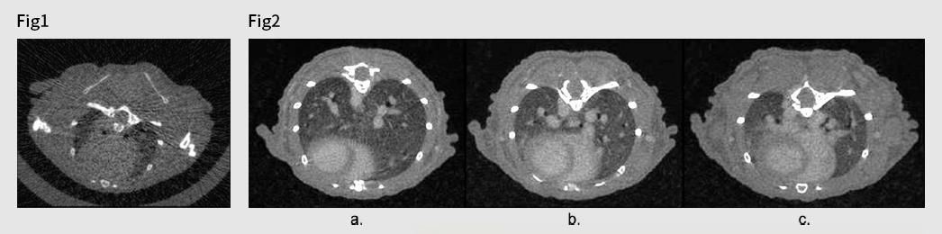
Fig1: 造影剤を使用せず撮影(横断面)。胸腔や心室では軟部組織のコントラストが悪い。
Fig2: Fenestra VC, iv投与2時間後のマウス、横断面像(Scanco Viva CT40撮影)
画像aおよびbは、心臓の両心室と胸腔内のいくつかの主要な血管を確認できる。画像cでは、両心室と心臓底部付近の胸部血管系も確認することができる。
(詳細:![]() Imaging Vascular Anatomy in Nude Mice Harboring Lung Carcinoma in Flank)
Imaging Vascular Anatomy in Nude Mice Harboring Lung Carcinoma in Flank)
| 製品名 | 容量 | 製品コード | 価格(税抜) |
|---|---|---|---|
| Fenestra VC | 2.5mL | VC-131 | 120,000 |
資料:Poster Presentations
- Micro-CT Contrast Agents for Detection of Murine Non-Alcoholic Fatty Liver Disease
- Micro-CT Based Quantification of Mouse Brain Vasculature: The Effects of Acquisition Technique and Contrast Material
- Novel microCT Imaging Techniques for in vivo Quantification of Vascular Volume in Murine Tumor Models
- In Vivo MicroCT Imaging Characteristics of a Long-Acting Blood-Pool Agent in Normal and Tumor-Bearing Mice
- Statistical Tracking of Murine Liver Metastases using a Hepatocyte-Selective Contrast Agent in Conjunction with MicroCT
- Early Detection of Liver Tumors in Mice using MicroCT and a Hepatocyte-selective Contrast Agent
- Effects of Administration Dose, Species and Strain Variations on MicroCT Imaging Characteristics of a Long-Acting Blood-Pool Agent inNormal Rodents
- Effects of Dose, Sex, and Species Variations on Imaging Efficacy of a Hepatocyte-Selective Contrast Agent in Normal Rodents
- MicroCT Evaluation of a Long-Lasting Blood Pool and Hepatobiliary Contrast Agents Following Multiple Intravenous Injections in NormalMouse Models
- Effects of Administration Routes on MicroCT Imaging Characteristics of a Long-Acting Vascular Contrast Agent in a Murine Model